X-ray tomography uses the ability of X-ray radiation to penetrate objects. On the way through an object, part of the impinging radiation is absorbed. The longer the radiographic length of the object, the less radiation escapes from the opposite side. The absorption also depends on the material. An X-ray detector (sensor) captures the escaping X-ray radiation as a two-dimensional radiographic image. At detector sizes of approximately 50 mm to 400 mm, a large portion of the measured object can be captured in a single image.

In order to use tomography on an object, several hundred two-dimensional radiographic images are made in sequence, with the measured object in various rotated positions (Fig. 35a). The object is located on a rotating table for this purpose, which is gradually rotated step by step. The three-dimensional information about the measured object contained in this series of images is extracted using a suitable mathematical process and is made available as a “voxel image”. Each voxel (volume pixel) embodies the X-ray absorption by the measured object for a defined location in the measured volume. Similar to two-dimensional image processing, the actual measured points are calculated from the voxel data using a suitable threshold process.

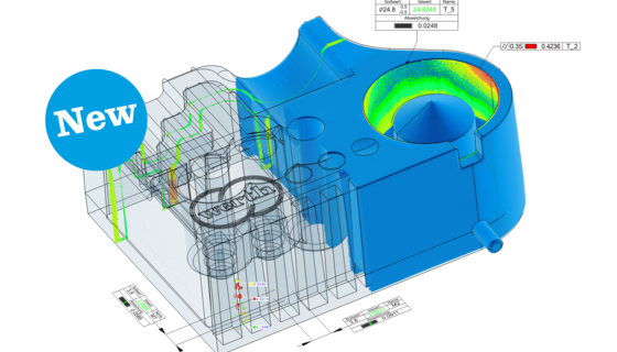
The sensors currently used capture up to 4 million image points. Typically, several hundred thousand to a few million measurement points are derived in the measured volume. These points are distributed evenly across the surface of the part being measured. Geometries in the interior of the measured object, such as hollow cavities or undercuts, are also captured. The measurement points can be evaluated using the familiar methods of coordinate measuring technology.

Similar to measurement using image processing, it is possible to change the magnification using tomography (Fig. 35b) in order to capture small parts with higher magnification, or larger parts completely with lower magnification. To do this, either the measured object is positioned in the radiation path or the X-ray components (X-ray source and detector) are moved in an axial direction relative to the measured object.

In some cases, the size of the sensor or the number of pixels available is still not enough to meet the requirements for the measuring task. In such cases, several images are stitched together (Fig. 35c) by moving the rotary table with the measured object relative to the X-ray components. Reconstruction of the voxel volume image is then accomplished on the basis of the stitched 2-D radiographic images.