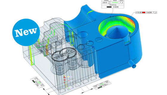
X-ray tomography uses the ability of X-ray radiation to penetrate objects. An X-ray tube can be considered almost a point source of X- rays. The X-rays pass through the measured object to reach the X-ray sensor. On the way through the object, a part of the radiation is absorbed. The longer the penetrated length of the object, the less radiation escapes from the opposite side of the object. The absorption also depends on the type of material.  This process is analogous to the creation of a shadow image of a partially transparent object by illuminating it with a point source of light. The brightness levels in the X-ray image correspond to the transparency of the penetrated areas and thus depend on their material density. The cone-shaped X-ray beam produces two- dimensional radiographic images of the object. The X-ray tomography sensor creates these images in an analogous manner to the image sensor in a digital camera. It provides the images in digital form for further analysis. In order to use tomography on an object, several hundred to a few thousand of such two-dimensional radiographic images are made in sequence.  The sequence is made with the measured object in numerous accurately known rotated positions.  The three-dimensional information about the measured object is contained in the digital image sequence thus generated. Using suitable mathematical methods, a volume model that describes the entire geometry and material composition of the work piece can be calculated from this sequence.  Due to the beam shape, this process is called cone beam tomography.