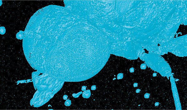


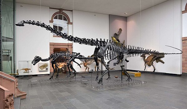
Since its foundation in 1817, the Senckenberg Gesellschaft für Naturforschung has been researching the development of the earth and the importance of biodiversity. The Senckenberg Nature Museum at the main site in Frankfurt am Main is one of the largest natural history museums in Europe. The Senckenberg's natural history collections – are the most extensive in Germany with around 41 million specimens –. As "archives of life", they form an indispensable basis for research into our living and non-living environment. Many of these collections can already be researched online and used all over the world. Digital technologies are crucial to making scientific collections accessible to everyone. This is because the way in which collection material is used has constantly evolved. Technical progress and new research methods make it possible to extract more and more information from objects. For the highly accurate digitization of the unique research objects, a TomoScope® XS Plus with a new sub-microfocus tube was jointly funded by the HMWK (ERDF) and the Senckenberg Gesellschaft für Naturforschung's own funds (including SOSA).
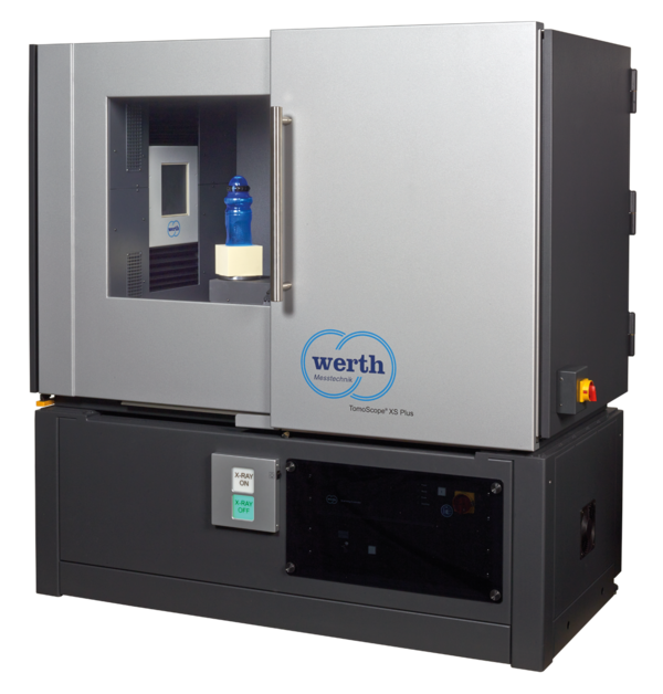
2017 Werth Messtechnik presented the TomoScope® XS, the first compact device with X-ray computed tomography for fast measurements with high structural resolution. Thanks to the innovative tube design, the maintenance interval for such machines can be extended to 12 months for the first time. With the first TomoScope® XS Plus – at that time still with 130 kV or 160 kV maximum voltage –, the measuring volume was quadrupled. Today, the compact device is available with up to 200 kV tube voltage. The higher voltage also allows the device to be used for workpieces made of dense materials with long radiographic lengths that are difficult to penetrate.
Werth is now introducing this machine type with the first sub-microfocus tube in monoblock design and long-life components. Until now, sub-microfocus tubes have been particularly maintenance-intensive, resulting in long downtimes and high costs. The new tubes with up to 160 kV voltage allow high availability and significantly lower maintenance costs compared to conventional sub-microfocus tubes with a separate voltage generator.
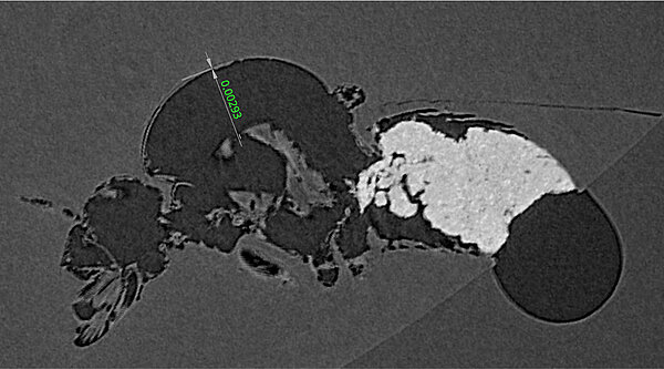
This is the first time that a sub-microfocus tube is available in this machine class. The maximum structural resolution in the 2D radiographic image is often specified as a characteristic. In practice, only the focal spot size of the X-ray source is relevant here; other influencing parameters are largely neglected. This allows very small numerical values of a few hundred nanometers to be specified. This value is around 0.8 μm for a TomoScope® XS Plus with the new sub-microfocus tube. However, the correct coordination with the other device components such as the rotary axis and detector is also crucial for structural resolution in the 3D volume. Temperature also has a major influence on resolutions in the limit range. For this reason, active temperature control to 20 °C ± 1 K is used inside the device. In addition, temperature-related drift is automatically corrected. All these measures make it possible to achieve a 3D volume structural resolution of around 1 μm, which is practically unsurpassed even by conventional, so-called "nanofocus systems".